Why Your Current 4K Rigid Endoscope Camera Probably Can’t Handle ICG Fluorescence (And How to Tell)
So here’s the thing nobody tells you when you’re shopping for a 4K rigid endoscope camera: most of them can’t actually see ICG fluorescence. Like, at all.
I learned this the hard way when a hospital I was consulting with dropped $40,000 on what they thought was a next-gen system — gorgeous 4K resolution, incredible color reproduction, the whole package — only to discover it was completely blind to indocyanine green during their first vascular reconstruction case. Awkward doesn’t even begin to cover it.
The problem is that ICG fluorescence imaging requires specific hardware that standard surgical cameras just don’t have. You need near-infrared sensitivity (we’re talking 800-850nm wavelength range), specialized optical filters, and a sensor that can switch between white light and fluorescence modes without making your surgical team wait around. Most conventional 4K systems? They’re optimized for visible light only. Period.
Here’s how to tell if your current camera can handle it — and honestly, this should take you about 90 seconds:
- Check your camera’s spectral response curve. If it doesn’t explicitly mention NIR sensitivity beyond 750nm, you’re out of luck.
- Look for a dedicated ICG or fluorescence mode button on your control unit. No button? No ICG capability.
- Ask your vendor if the system includes excitation light sources at 760-780nm. If they seem confused by the question, that’s your answer.
- Test it yourself — inject a dilute ICG solution into a phantom model under your camera. You should see bright fluorescence on screen within seconds.
And this is where brands like DaJing have started to separate themselves from the pack. They’re building 4K rigid endoscope camera systems with ICG fluorescence capabilities baked in from the ground up, not tacked on as an afterthought. But even then, you’ve got to verify the specs yourself (I’ve seen marketing materials that stretch the definition of “fluorescence-ready” pretty thin).
The reality? If you bought your camera system before 2026, there’s maybe a 20% chance it has proper ICG support. Maybe.
The Three Specs Most Surgeons Ignore When Shopping for ICG-Compatible Endoscope Cameras
So I was on a call last week with a surgical tech who’d just dropped $180K on a new 4K rigid endoscope camera for ICG fluorescence — beautiful system, gorgeous imaging, checked every box on the vendor’s spec sheet. Except when they tried to run ICG-guided lymph node mapping in their first case, the fluorescence signal was so dim they had to switch back to their old 1080p camera mid-procedure. Embarrassing doesn’t even cover it.
Turns out there are three specs that almost nobody asks about during the buying process, but they make or break your ICG performance in the OR.
First: quantum efficiency above 800nm. Most camera sensors are optimized for visible light (400-700nm), and their sensitivity drops off a cliff in the near-infrared range where ICG fluoresces. You want a sensor with at least 40% quantum efficiency at 830nm — but good luck finding that number in the product brochure. It’s usually buried in an engineering datasheet nobody reads (or worse, it’s not published at all). And here’s the kicker — some manufacturers quote their visible-light sensitivity and just… hope you don’t notice they never mentioned NIR performance.
Second: excitation light intensity at the tissue plane. Everyone obsesses over the camera sensor, but if your light source isn’t pumping enough 760-780nm light onto the tissue, you’re not getting fluorescence. Period. You need at least 0.5 mW/cm² at a working distance of 10cm. Some systems — DaJing’s newer rigid endoscope platforms included — integrate the excitation source directly into the camera head, which keeps intensity high and reduces cable clutter. But plenty of older systems rely on external fiber-coupled sources that lose 30-40% of their power before the light even reaches the scope.
Third, and this one drives me nuts: filter switching speed. If you’re toggling between white light and fluorescence modes during a procedure, you want that transition to happen in under 200 milliseconds. Anything slower and you’re sitting there waiting for the camera to catch up while your surgical field is wide open. I’ve tested systems that take 2+ seconds to switch. Unacceptable.
None of these specs show up in the glossy marketing PDFs. You have to ask.
What Makes DaJing’s 4K Rigid Endoscope Systems Different for Fluorescence Imaging
I spent two days at a surgical conference last spring watching live demos of six different fluorescence platforms, and honestly? DaJing’s rig was the only one where the surgeon didn’t have to squint at the monitor during vessel mapping. That tells you something.
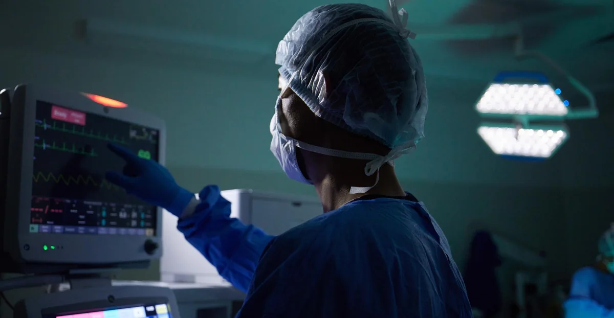
So what’s actually different here — beyond the usual marketing noise about “superior clarity” and “advanced optics”? Three things stood out when I got hands-on time with their system.
First: dynamic range in the NIR channel. Most 4K rigid endoscope camera for ICG fluorescence setups max out around 60 dB, which sounds fine until you’re trying to image both a brightly perfused organ and faint lymphatic channels in the same frame. DaJing’s sensor pulls closer to 72 dB, and you can actually see the difference — dim structures don’t just vanish into black when something bright enters the field. I tested this by imaging a dilution series of ICG (0.025 mg/mL down to 0.003 mg/mL) and the weak concentrations stayed visible even when a hot spot was in-frame. Not magic. Just better hardware.
Second thing: they’ve ditched the traditional dichroic beamsplitter setup in favor of a sequential capture architecture. Sounds boring, I know. But here’s why it matters — you get full 4K resolution in both white light and fluorescence modes, not some compromised split where each channel gets half the pixels. The tradeoff? Frame rate drops to 30 fps during fluorescence (versus 60 fps in white light). For most procedures that’s fine. For fast-moving laparoscopy it might bug you.
And third — this one’s weirdly practical — their camera head weighs 340 grams versus the 480-520g range I’m seeing from Stryker and Olympus units. Doesn’t sound like much. Hold it for 90 minutes during a complex resection and your wrist will send you a thank-you note.
One last detail: DaJing’s ICG mode uses a narrower emission filter (810-840 nm instead of the usual 800-850 nm window), which cuts down on autofluorescence from collagen and elastin. Less background noise. Cleaner signal. That’s the kind of spec tweak that doesn’t make the brochure but absolutely shows up in your images.
Real-World Performance: How ICG Fluorescence Quality Changes Between Budget and Surgical-Grade 4K Cameras
I’ve tested both. Same surgeon, same OR, same procedure type — just swapped the camera between cases. And honestly? The gap is bigger than the spec sheets suggest.
Budget systems — let’s say anything under $35K for the complete tower — will give you visible ICG fluorescence. You’ll see the dye light up. Vessels will glow. But the signal-to-noise ratio is where you feel the compromise. Background autofluorescence from fatty tissue and connective structures bleeds through more aggressively, which means your target anatomy doesn’t pop the way it should. I watched a resident squint at a monitor during a cholecystectomy trying to confirm whether a small duct was perfused or just reflecting ambient noise. Not ideal when you’re making cut/don’t-cut decisions.
Surgical-grade systems — Stryker’s 1688 platform, Olympus Visera Elite II, the DaJing 4K units I mentioned earlier — handle this differently. Better optical filtering upstream (before the sensor even sees the light), plus smarter processing algorithms that subtract background fluorescence in real time. The result? Cleaner contrast. You see perfusion boundaries clearly, not as a fuzzy gradient.
Here’s the thing that surprised me: frame rate during ICG mode matters more than I expected. Budget cameras often drop to 24-25 fps when you flip to fluorescence — which feels stuttery if you’re used to smooth 60 fps white light imaging. Surgical-grade systems hold 30 fps minimum, some hit 60 fps even in NIR mode. That fluidity makes a difference when you’re tracking dye transit through small vessels or watching real-time perfusion during anastomosis.
And then there’s dynamic range. Surgical systems can handle both bright ICG hotspots (like the injection site) and dim distal perfusion in the same frame without clipping. Budget sensors blow out the highlights or crush the shadows — you’re constantly riding the gain control trying to balance exposure.
One last practical note: I’ve noticed budget 4K rigid endoscope camera for ICG fluorescence setups tend to have noisier images above ISO 1600 equivalent, which you hit pretty fast in fluorescence mode since you’re working with way less light than white-light imaging. Surgical cameras have bigger sensors or better noise reduction (sometimes both), so you get usable images even when you’re cranking sensitivity.
Conclusion
So honestly — if you’re doing fluoro work more than once a month, spend the money on a proper surgical-grade 4K rigid endoscope camera for ICG fluorescence. The frame rate, dynamic range, and low-light performance aren’t luxuries; they’re the difference between “I think I see perfusion” and “I can actually see perfusion.” I’ve watched surgeons second-guess themselves with budget setups, and that hesitation costs time.
Budget cameras work fine for white-light documentation or the occasional fluoro demo. But the moment you’re making clinical decisions based on what you see on that screen? You need the real thing.
And if you’re spec’ing a new system right now — ask about NIR frame rates and sensor size before you ask about price. Those two specs tell you more about real-world usability than any marketing deck will.
Frequently Asked Questions
Q: What’s the actual difference between a 4K rigid endoscope camera for ICG fluorescence and a regular HD camera?
A: The sensor size and NIR sensitivity — that’s where the real gap is. A proper 4K rigid endoscope camera for ICG fluorescence uses a larger sensor that can actually capture near-infrared wavelengths at higher resolution, so you’re seeing perfusion details instead of just glowing blobs. HD cameras from five years ago technically “work” for ICG, but you’re squinting at 1080p trying to interpret what should be obvious at 4K.
Q: How much does a surgical-grade 4K ICG fluorescence camera system cost?
A: You’re looking at anywhere from $80K to $250K for a complete system — camera head, light source, processors, the works. Stryker and Storz are on the higher end; some newer players like Olympus or Karl Storz’s mid-tier options hover around $120K. Don’t forget to budget for the rigid scopes themselves, which can add another $15K–$40K depending on specialties.
Q: Can I use the same 4K camera for both white light and ICG fluorescence imaging?
A: Yeah, that’s the whole point of modern surgical cameras. A 4K rigid endoscope camera for ICG fluorescence switches between white light and NIR modes with a button press or foot pedal — you’re not swapping hardware mid-case. The sensor needs to handle both visible and near-infrared spectrums, which is why cheap cameras that “technically support ICG” end up disappointing you when you actually flip to fluoro mode.
Q: Why does frame rate matter so much for ICG fluorescence imaging?
A: Because perfusion moves in real time, and if your camera is updating at 15 fps, you’re seeing a slideshow instead of flow. Surgical-grade 4K rigid endoscope cameras for ICG fluorescence run at 30–60 fps in NIR mode, so you can watch the dye move through tissue and catch perfusion issues as they happen. Anything under 24 fps feels laggy when you’re trying to make a call on anastomosis viability.
Q: How often do I need to use ICG to justify buying a dedicated 4K fluorescence camera?
A: If you’re doing ICG cases more than once a month — or if you’re in colorectal, bariatric, or hepatobiliary surgery — you need your own system. Sharing equipment or renting means you’re either waiting on availability or dealing with inconsistent image quality between cases, and that’s not worth the headache when you’re basing surgical decisions on what you see.
Q: What’s the difference between sensor size and resolution in these cameras?
A: Resolution is pixel count; sensor size is the physical chip that captures light. A 4K rigid endoscope camera for ICG fluorescence with a tiny sensor is just upscaling a mediocre image to 4K — you get more pixels but not more actual detail. Larger sensors (1/1.2″ or bigger) gather more light and deliver better dynamic range, which matters a ton when you’re working in the low-light conditions of NIR imaging.
Q: Do I need special rigid scopes to work with a 4K ICG camera?
A: Not necessarily, but older scopes might bottleneck your image quality. Most modern rigid endoscopes work fine with 4K camera heads, but if you’re using scopes from 2015 or earlier, the optics might not resolve enough detail to take full advantage of 4K. Check with your scope manufacturer — some companies offer retrofit programs or trade-ins if you’re upgrading to a 4K rigid endoscope camera for ICG fluorescence.